Kinematics of the Normal Knee Based on Videofluoroscopy
Description
Total arthroplasty of the knee aims to relieve patient pain and improve functionality of the joint. To maintain sufficient range of motion, but also not overload the surrounding soft tissue structures, reproduction of tibio-femoral kinematics of the healthy knee is thought to be beneficial. For an evaluation of current TKA designs, as well as for improvement and the development of newer products, accurate knowledge of the three-dimensional kinematics of the normal knee is therefore crucial.
The movement of the normal tibio-femoral joint has been extensively investigated in cadaver studies, using MRI or CT as well as by bone-pins, RSA and videofluoroscopy. In more traditional studies using MRI, during flexion the medial condyle, described by the centre of the medial sphere, translates no more than ±1.5mm, whereas the lateral condyle translates posteriorly about 15mm by a combination of rolling and sliding. In contrast to the latter studies recent publications suggest that during the stance phase of gait, as well as running the joint center of rotation is predominantly on the lateral side, thus knee movement described resembles a lateral pivot movement. There are a variety of different methods used to describe tibio-femoral motion, complicating the comparison between different studies. Many studies use a circular axis fixed in the femur, other describe motion by means of an epicondylar axis, by using an axis joining the lowest points on the femoral condyles or an axis joining the contact points. The definition of the axes used to describe tibio-femoral motion therefore seems to be important, but selection of the “correct” axis remains controversial.
It is clear that tibio-femoral motion in the natural knee during normal activities of daily living remains controversially discussed, while it remains unknown whether the loading conditions or the analysis technique is responsible for this discrepancy.
Goal
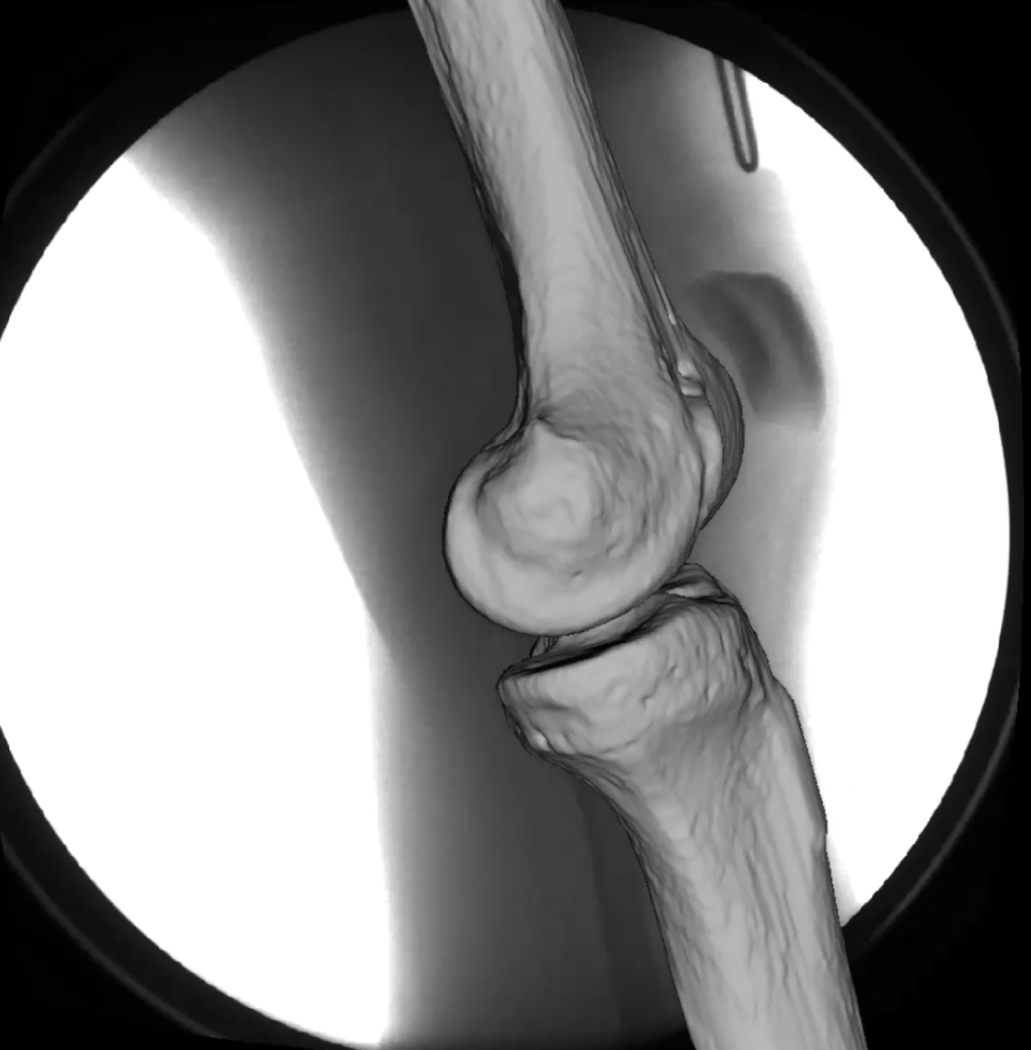
This project aims to investigate the normal knee during activities of daily life by means of videofluoroscopy and an automated moving fluoroscope developed at the ETH Zurich. It further includes 2D/3D registration based on CT data, as well as the investigation of the role of anatomical landmarks (circular axis, epicondylar axis) vs functional axes in the determination of tibio-femoral kinematics.
Contact
Head of Dep. of Health Sciences and Technology / Deputy head of Institute for Biomechanics
Institut für Biomechanik
Gloriastrasse 37/ 39
8092
Zürich
Switzerland
