From back shape to spine: radiation-free assessment of posture, motion and pathologies of the spine
Background
Spinal disorders and bad posture are quite common and difficult to access and monitor accurately. The assessment and monitoring of the spine involves radiographic assessments of the entire upper body at multiple time points, resulting in cumulative exposure to radiation. 3D optical systems provide an alternative to assess the 3D spinal curvature without ionising radiation.
Goal
The goal of this project is to develop and clinically validate algorithms to generate dynamic 3D models of the human spine. These algorithms are solely based on the 3D back surface and utilize statistical shape modelling and machine learning.
Method
Within a clinical trial together with our partner, the Balgrist University Hospital, we collect 3D captures of back surfaces of adolescent idiopathic scoliosis patients together with the corresponding x-ray images. Based on this dataset we develop algorithms to generate dynamic 3D models of the human spine. The back surfaces are captured with different methods and different 3D capturing systems. Various algorithms based on the asymmetry of the back shape are developed, improved and compared to the results from X-ray images.
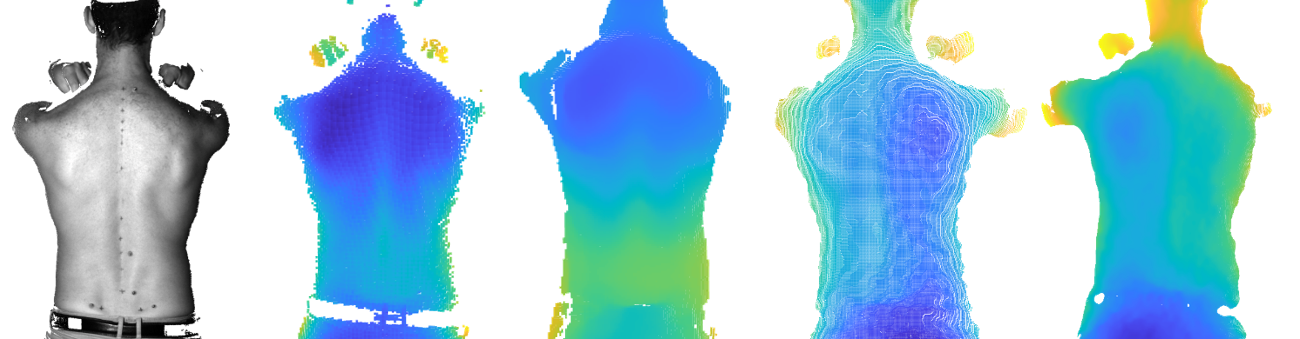
Validation of predicted spinal alignment from the 3D back shape with the corresponding X-ray images
Contact
Professur f. Bewegungsbiomechanik
Gloriastrasse 37/ 39
8092
Zürich
Switzerland

Funding
This project was funded by the Innosuisse Translational Research Funding (47195.1 IP-LS).
Collaboration
This project was done in collaboration with Bern University of Applied Sciences and Balgrist University Hospital.